Pet Owner’s Guide 101: Ocular Proptosis in Dogs

Ocular proptosis is the protrusion of the eye from its socket

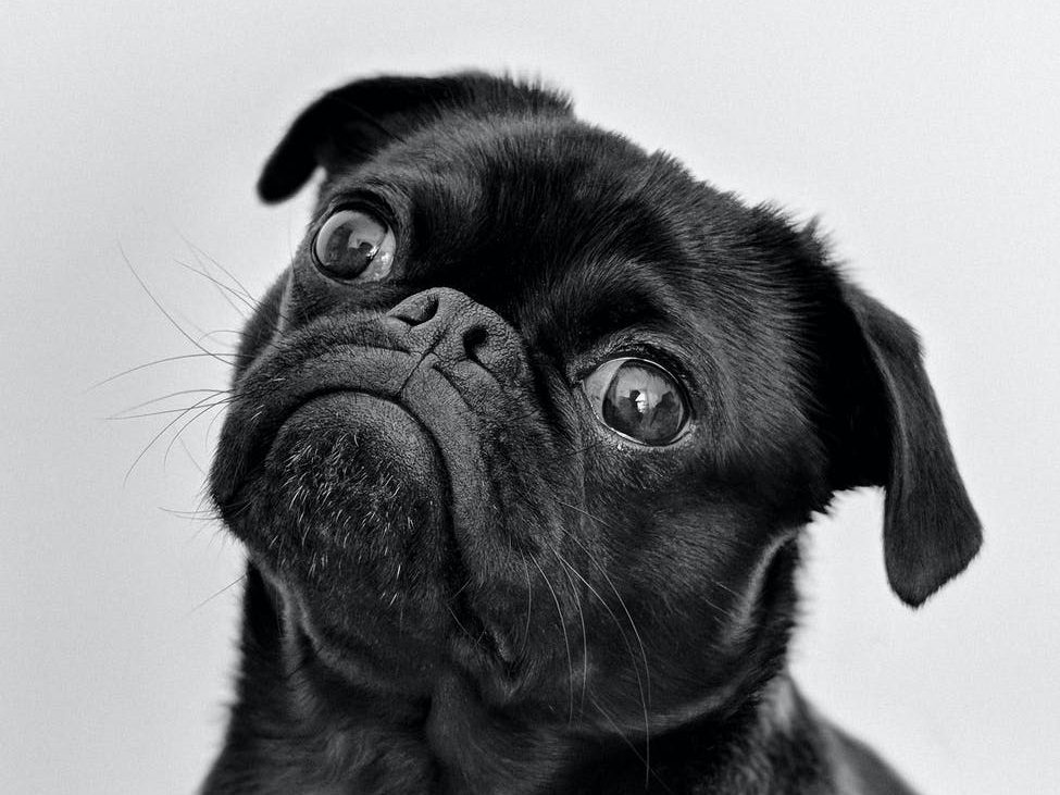
Brachycephalic breeds of dog, such as shih tzu’s, pugs, bulldogs, and pekingese dogs are well-represented in cases of ocular proptosis. This is due to their facial structure wherein their eye sockets are shallow, making it easy for the dog’s eyes to bulge out.

Left: Ocular proptosis in a brachycephalic dog.
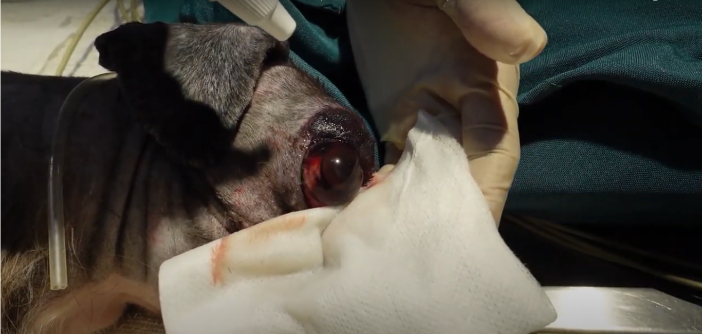
Right: Healthy eye after treatment.

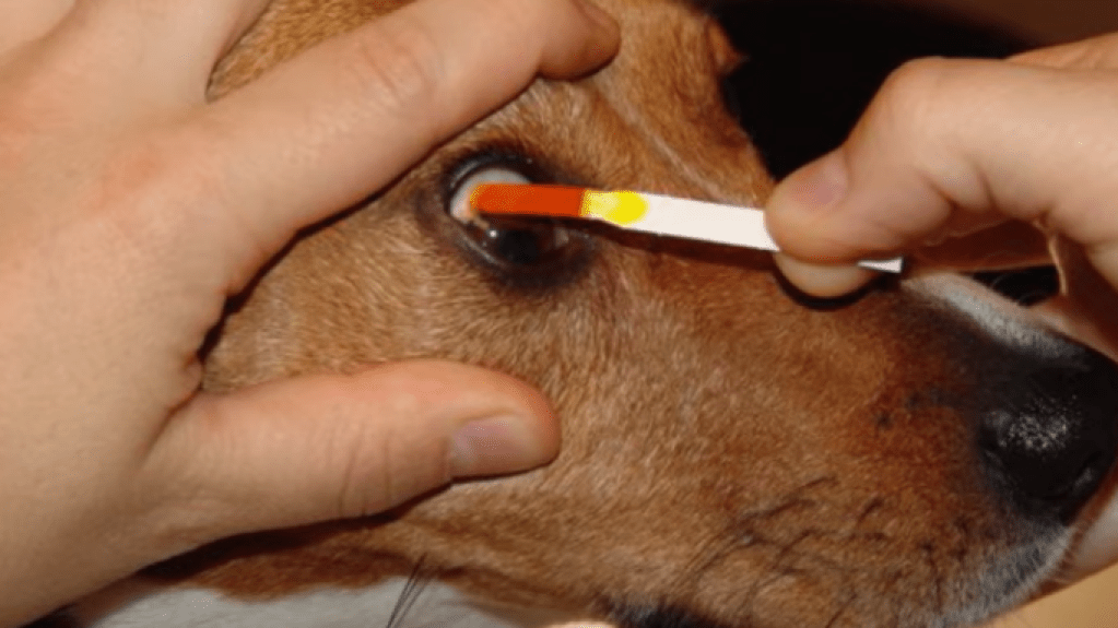
Ocular proptosis should be treated as an emergency. The mere dryness of the overexposed eye (corneal desiccation) can lead to corneal ulceration. There is also high risk of blindness, among other complications.

Click here for first aid.

Table of Contents

Symptoms

Proptosis is relatively easy to detect through physical inspection. The affected eye may be completely protruding or only slightly.

If a pet owner is unsure if the eye is indeed bulging, here are things to look out for:

  • Uneven eyes (affected eye is protruding)
  • Strabismus (cross-eye)
  • Corneal desiccation (dry eye) when at least 1 hour has elapsed
  • Conjunctivitis, reddish blood vessels in eye
Side view of a dog’s eye affected by traumatic proptosis.

It also helps to view the dog’s eye from the side or from the top of their head, as this may make the difference in the size of the two eyes more apparent than if they are viewed upfront.

Causes

The main cause of proptosis is trauma such as those from falls and dog fights. 

This may not only be trauma near the eyes, but also on certain parts of the body such as the neck. 

When it comes to brachycephalic breeds with protruding eyes such as pugs and bulldogs, even pressure on the neck from a collar may result in ocular proptosis due to their facial structure having shallow eye sockets.

Although not as common as trauma, pressure from tumors at the back of the eye may also lead to it bulging out.

First Aid

Ocular proptosis requires immediate attention. If your dog or small animal’s eye protrudes from its socket, follow these steps:

  1. Find a clean piece of cloth. 
  2. Dampen it.
  3. Gently cover the animal’s eye. 
  4. Call your vet and bring the animal to the clinic.

If the dog hasn’t eaten yet, that’s fine. Give them water, or if they’re overdue for feeding, limit the amount of food you give; although the procedure will be on their eye, it’s likely that they’ll need their stomach empty in preparation for surgery.

Treatment Methods

Treatment depends on the severity of the proptosis, as well as the pet owner’s capacity for long-term care.

The patient is prepped for surgery. Visible are the swollen eyelids.

Eyeball Replacement

Eyeball replacement, or globe replacement, involves helping the eyeball re-align with the eye socket through temporary tarsorrhaphy. 

Tarsorrhaphy is a surgical procedure wherein the eyelids are sewn together. In temporary tarsorrhaphy, this is done to protect the eye and facilitate recovery until the eye is healed enough to be un-sewn.

Eyeball replacement is generally more preferred over irreversible enucleation.

Eyeball replacement is apt for patient dogs who have mild cases of ocular proptosis and whose eyeball can be salvaged and is aesthetically satisfactory to the pet owner.
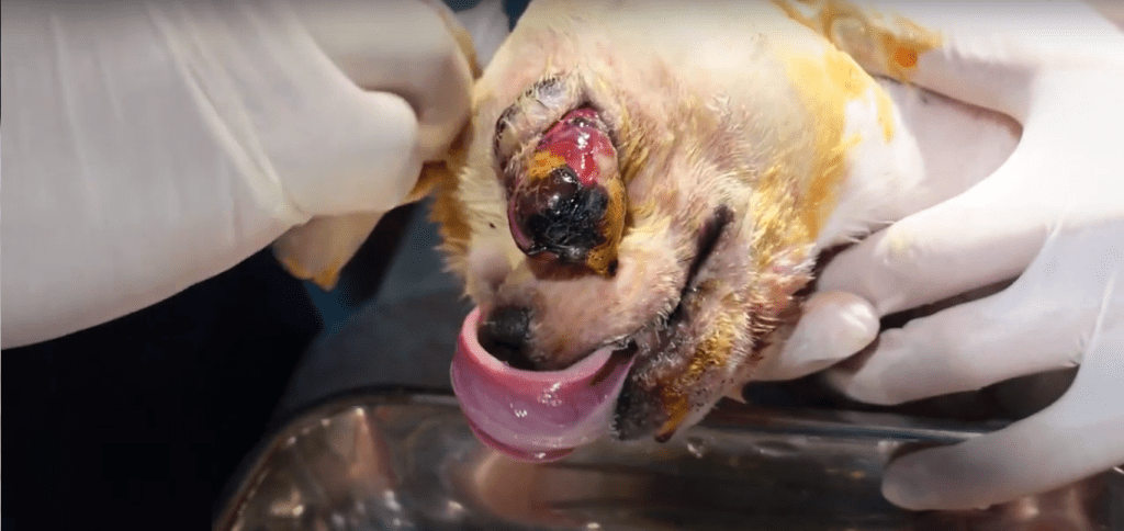
Enucleation

Enucleation or complete removal of the dog’s eye is considered when all other options have been exhausted. 

If the patient is a strong candidate for enucleation, enucleation may also be the chosen procedure, without having the animal first undergo eyeball replacement.

A patient with proptosis is a strong candidate for enucleation if the optic nerves of the affected eye are broken, or if there is hemorrhage, infection, tumor, or advanced glaucoma.

For pet owners unable or hesitant to provide long-term care, enucleation is also considered.

Recovery

After surgery, the patient recovering from ocular proptosis may experience strabismus or cross eye. This may last 3 to 6 months, sometimes 9 depending on different factors, after which the eye may return to normal.

Said factors affecting prognosis are:

  • duration of corneal exposure
  • breed
  • damage to the eye and its surroundings

Case

From the Philippines? Check out our case vlogs. Same veterinary content, in audiovisual format.

Ocular Proptosis FAQs

What breeds are prone to ocular proptosis?

Breeds with protruding eyes are prone to proptosis. These include pugs, bulldogs, shih-tzu’s, and Pekingese.

Does my pug have proptosis? I can’t tell because his eyes are always bulging.

A simple way to check is to compare one eye to the other. It also helps to observe the eye from the side or from the top of the dog’s head, rather than from the front. See also: symptoms.

My dog’s breed is prone to ocular proptosis. How do I prevent him from getting it?

Review the causes. Here is a non-exhaustive list of preventive measures:

  • Neuter your pet. In prone breeds, proptosis may result from tugs at the collar or leash, such as when preventing them from entering into a dog fight.
  • Socialize your pet with other dogs. As with neutering, the goal here is to make your dog less prone to aggression, so there will be no need to pull them from the collar and less sources of trauma.
  • Beware of other dogs. If you can, pick up your dog and keep them away from unfamiliar dogs that may initiate the dog fight.

Will my dog become blind from proptosis?

It depends on the severity of the case.

My dog’s eye is protruding. Can it go back in?

A vet may professionally restore the eye. However, this depends on the its state.

See also: Will my dog lose his eye?

Will my dog lose his eye from traumatic proptosis?

It depends on the state of his eye. 

Damaged optic nerves, hemorrhage, infection, tumor, and advanced glaucoma are some of the considerations that go into deciding whether enucleation is an appropriate treatment method.

If the eye is in a good state, it may simply be replaced. See Treatment Methods.

Pet Owner’s Guide 101: Bladder Stones in Dogs

Bladder stones are fairly known health concerns in humans as well as in dogs. Their most common symptoms are blood in urine and painful urination, or hematuria and dysuria.

Despite “bladder stones” being easy to pronounce and recognizable in name, suspecting your dog might have bladder stones thanks to these clear symptoms isn’t enough to figure out the right treatment method. There are multiple types of bladder stones, with distinct causes and corresponding treatment.

In this article, join us as we discuss two common types of bladder stones, their causes, how to treat them, and how to prevent them.

From the Philippines? Check out our case vlogs. Same veterinary content, in audiovisual format.

Table of Contents

Symptoms

As mentioned, hematuria (blood in urine) and dysuria (painful urination) are the main symptoms of bladder stones.

Secondary symptoms may be urination in unusual places and unusually frequent urination.

Swollen blood vessels of the bladder. If urinary obstruction from bladder stones is left untreated, the bladder may rupture.

Types and Causes

There are several types of bladder stones. Why is it important to mention the type of bladder stone?

This is because, in order to know the right treatment and prevention methods for your pet, proper identification of the type of bladder stone is a must.

For the purpose of this article, two common types of bladder stones will be discussed: struvite stones and calcium oxalate stones.

Struvite stones

At low levels, struvites and struvite crystals are not a major cause for concern. What becomes a problem is when they form larger struvite stones and risk urinary obstruction.

Struvites are formed when urine becomes alkaline. Dog urine is usually slightly acidic. Since urine turns alkaline when there is urinary tract infection or UTI, struvite stones are regarded as infection stones.

Calcium oxalate stones

Calcium oxalate stones, on the other hand, form regardless of UTI. They have less to do with bacterial infection and more to do with genetic risk factors.

Unlike struvite stones, existing calcium oxalate stones are not dissolved by changing your pet’s diet to a less alkaline one at the last minute. When it comes to calcium oxalate stones, the goal is to prevent their formation.

Complications

Apart from painful urination, leaving bladder stones untreated may cause further complications. It leads to urinary obstruction, especially in male dogs. This has the potential to be life-threatening; if neglected, the bladder may rupture.

Treatment Methods

Dietary Dissolution

Dietary dissolution is a treatment method wherein a dog’s diet is changed to be less alkaline. It is used for struvite stones, which again, are formed due to bacterial infections.

Because it only changes the acidity of urine, this treatment method does not work for calcium oxalate stones.

Dietary dissolution is a good choice, however, for struvite stones. The duration of treatment is proportional to the size and number of struvite stones present.

Surgical Procedure

Surgical removal of bladder stones is an option for both struvite and calcium oxalate stones

It is a straightforward procedure where the bladder stones are manually taken out. This removal of the bladder stones is called cystotomy.

Cystotomy performed on a dog. A large bladder stone is taken out.

Urethrostomy, a distinct surgical procedure, is meant to address future formation of bladder stones. It involves creating a passageway for bladder stones to exit the body. This treatment method is chosen when eventual formation of new bladder stones is probable, such as in the case of calcium oxalate stones in dogs with genetic risk factors.

Patient dog being prepared for the surgery.

Lithotripsy

In some countries, lithotripsy may be used to treat bladder stones. Lithotripsy uses ultrasound waves to dissolve the bladder stones. It is a non-invasive treatment method using advanced technology and is not widely available in all countries.

Prevention

Prevention methods are crucial when it comes to preventing the formation of bladder stones and addressing them in time if they do form. These measures are great for different types of bladder stones, but especially with calcium oxalate stones that are genetic.

These prevention measures include:

  • Maintaining a diet with a balanced pH level (more acidic than alkaline)
  • Encouraging your dogs to drink plenty of water
  • Feeding dogs with wet canned food if they dislike drinking water often
  • Booking regular check-up for dogs with risk factors

Cases

We have cases for both struvite stones and calcium oxalate stones. Both cases are documented in this video. For the purpose of this article, however, we’ll be discussing the calcium oxalate case.

What’s interesting about our patient dog who had calcium oxalate bladder stones is that he was brought in due to skin problems.

It was definitely a separate issue from his bladder stones. We learned of the bladder stones when we noticed that the dog experienced hematuria.

We conducted tests including ultrasound to determine the type of bladder stone, as some types may not be detected by an x-ray machine and we wanted to provide the appropriate treatment.

Upon confirming that the stones inside the bladder of the patient bulldog were calcium oxalates, it was deduced that cystotomy was needed to remove the existing stones before they formed into larger ones, as well as urethrostomy in preparation for the likely formation of newer stones.

Bladder Stones FAQs

“I don’t want to have my dog undergo surgery. What can I feed my dog?”

Food that are more acidic may help balance the pH level of dogs with alkaline urine and prevent the formation of struvite-type bladder stones.

However, if you suspect that your dog already has bladder stones, please consult a vet for dietary recommendations.

“My dog has bladder stones again. What should I do?”

Have the type of bladder stone determined by a vet, as different types of bladder stones have different causes and thus, different treatment methods.

Calcium oxalate bladder stones, for example, are not dissolved by changing the diet, which works for struvite bladder stones.

It is also possible for one dog to form more than one type of bladder stone in their lifespan.

Pet Owner’s Guide 101: Corneal Ulcers in Dogs

Corneal ulceration is painful. It may also lead to blindness, and if left untreated, necessitate the removal of the eye.

Before catastrophizing into extremes, however, it’s important to spot corneal ulceration, its different levels of severity and mildness, what treatment methods there are, when those treatments are considered, and how to avoid the more serious of corneal ulcerations.

Speak Filipino? Check out our YouTube case vlogs. Same veterinary content, in audiovisual format.

Table of Contents

Symptoms

As the cornea is transparent, corneal ulcerations may not be visible to the naked eye. In order to see them, veterinarians add a fluorescent stain to the dog’s eye.

However, there are signs of corneal ulcerations that are more noticeable and that pet owners can look out for even at home.

Fluorescent stain is added by the vet to the dog’s eye to check if there is corneal ulceration.

Excessive watering of the eyes

Natural watering of the eyes, or lacrimation, is important in keeping your dog’s eye moist. 

If your dog’s eyes are producing tears excessively or more than usual, however, it may then be a symptom of corneal ulceration.

What does excessive lacrimation look like?

Excessive lacrimation may produce tear stains on your dog’s face. You can observe this by checking whether or not the fur under your dog’s eyes is wet.

If the fur under their eyes is always wet but the dog is not newly bathed, or only that part of their face is wet, then the lacrimation is already excessive.

Conjunctivitis in a dog’s eye.

Conjunctivitis

Just as in humans, conjunctivitis in dogs may be observed from the white part of the eyes turning reddish.

Whitish opacity

When it comes to injury on other parts of the body, inflammation may look pinkish. In the eyes, the inflammation manifests in the form of an opaque white film.

Eye twitching

Blepharospasm or eye twitching is another symptom of corneal ulceration.

This blinking and twitching is involuntary. As amusing as it may be to think that our dogs wink at us, involuntary eye twitching is their way of telling us that they might need a little check-up. 

Risk Factors

Breeds prone to corneal ulcerations include: pugs, bulldogs, and shih tzu’s. These brachycephalic breeds have prominent eyes.

Causes

The primary cause of corneal ulceration in dogs is trauma. This trauma usually comes from dog fights and play, though it may also occur from the dog’s rubbing their head against surfaces of door mats and kennels. 

Including trauma, causes of corneal ulcers in dogs are:

  1. Trauma or physical injury
  2. Genetic factors, breed of dog
  3. Keratoconjunctivitis sicca (KCS)
  4. Cushing syndrome
  5. Infections (less common in dogs than in cats)

If the latter causes of corneal ulceration sound intimidating, do not worry; consult a vet so they may rule out the accurate cause and provide the best treatment option for your pet.

Later in this article, we’ll discuss one corneal ulceration case we received in our Clinic, and how our veterinarians arrived at both the diagnosis and appropriate treatment method. 

Treatment Methods

Before determining the treatment appropriate to your dog’s needs, vets first asses the state of your dog’s eye. The extent of damage on the eye determines which treatment method is best.

One treatment method for corneal ulcerations in dogs is to temporarily close the eyelids of the affected eye in order to protect it and allow it to heal. 

In humans, eye patches are used. Given the impracticality of attaching an eye patch to even the most well-trained dogs, their eyelid is used to cover the eye instead. Covering the eye means conducting an operation in which the eyelids are sewn temporarily.

Upon recovery, the dog’s eyelids are un-sewn by the vet, and prescribed medicinal eye drops.

For some cases, dead tissue may first need to be removed from the cornea and replaced. This is called corneal grafting. The dead tissue is removed because it can no longer heal. Corneal scarring may be addressed with laser surgery.

Removal of the eye

The eye heals. Enucleation or removal of the dog’s eye is unnecessary unless the damage is major. 

The severity of damage corresponds to the parts of the eye affected.

Parts of the eye

Regarding corneal ulcerations, parts of the eye we are concerned with are the epithelium, stroma, and descemet’s membrane

Among the three, the epithelium is the outermost layer of the eye. When only the epithelium is damaged, this may be called a corneal abrasion. Corneal abrasion or corneal erosion is superficial trauma to the eye. Such shallow cuts heal in 3 to 5 days.

Next, we have the stroma. Compared to the epithelium, the stroma is further inside the interior of the eye. When the damage reaches the stroma, it already requires the aid of a vet.

Finally, we have the Descemet’s membrane. The Descemet’s membrane is the innermost layer among the three. This membrane is critical. Enucleation is considered when damage penetrates this layer. 

In order to avoid a descemetocele developing and necessitating enucleation, it is important not to leave corneal ulceration untreated. While the damage is still on the epithelium and stroma layers, have your pet receive medical attention from a vet.

Case

As previously mentioned, the primary cause of corneal ulceration is trauma. Trauma usually occurs in aggressive dog play and dog fights.

However, in a corneal ulceration case we handled in our Clinic, the dog did not have other dogs to play or fight with; neither were there cats.

The owners were puzzled that their dog’s eye was damaged. When we examined the dog, we found that he had another health concern: ear mites.

Our patient was teeming with ear mites, which caused him to keep scratching at his ears and face. It is likely that in the process of relieving himself from itching, he accidentally scratched his eye as well.

Fortunately, the corneal abrasion did not reach the Descemet’s membrane

Our senior veterinarian performed a minor operation on the dog’s eyelid, closing the conjunctival flap to protect the eye, and prescribed medicine to aid healing.

Because the owner brought the dog to the vet in a timely manner, the dog’s eye was saved from enucleation.

Puppy Eyes for Life

Photo by Charles on Pexels.com

Corneal Ulceration FAQs

“Will my dog’s eye be removed?”

Not all cases of corneal ulceration need to have the eye removed. It depends on the severity of the damage to the eye. For more information, see Treatment Methods.

“Can corneal ulceration cause blindness?”

If left untreated, the corneal rupture may progress to irreparable damage. Timely check-up and treatment can prevent this.

“Does corneal ulceration heal on its own?”

Corneal ulceration is damage to the eye that has reached a relatively deep layer called the stroma. Medical attention from a vet is needed, in order to prevent the ulceration from progressing.

Once the damage has progressed into an even deeper layer called the Descemet’s membrane, urgent medical attention is needed in order to avoid losing the entire eye. Consult a vet before this happens.

“How long do corneal abrasions heal in dogs?”

It may take 3 to 10 days for corneal abrasions to heal. Superficial corneal abrasions heal more quickly, while deeper corneal ulcerations take longer to heal. Corneal ulcerations left untreated may result in the damage progressing to the Descemet’s membrane, leading to the leaking of the descemetocele, at which point, enucleation or removal of the eye may be needed. Timely treatment, however, can prevent the need for enucleation.

“What’s the difference between corneal ulceration and corneal abrasion?”

Corneal ulceration may result from an abrasion in the cornea. If there is corneal ulceration, there is corneal abrasion.

“My dog keeps blinking. What does this mean?”

Frequent involuntary twitching of the eye could mean several things. If coupled with other symptoms such as excessive watering of the eyes or a white film on the eye, one possible case is corneal ulceration

It is best to consult a vet in person. However, this article on corneal ulcerations provides basic information to know.

“My dog’s eyes are always wet. Should I be concerned?”

This is called excessive lacrimation. Although it’s normal for your dog’s eyes to self-lubricate, excessive tears may be a symptom of a medical concern. Check for other symptoms and consult a vet.

If the symptoms include eye damage or eye twitching and blinking, you may read this article on a medical concern that has excessive lacrimation as a symptom

Our Clinic is also open for consultations.

“There’s a white film on my dog’s eye. What’s a good eye drop?”

If your pet has received a diagnosis from your vet, you may browse a catalog of products we carry.

Otherwise, do know that the white film may be a symptom of underlying medical concerns. It is best to consult a vet for advice and treatment best suited to your fur-baby’s needs.

If you need more information before going to the vet, you may read this article on corneal ulcerations, one medical concern that has the white film as its symptoms. Do consider the treatment best suited to your fur-baby’s needs, arrived at with proper diagnosis.